User-agent: Google Allow: A Simple Guide to Medical Conditions: August 2007

Search This Blog

Friday, August 31, 2007

A Simple Guide to Breast Cancer


A Simple Guide to Breast Cancer
--------------------------------------

What is Breast Cancer?
---------------------------

Breast cancer is a disease in which malignant (cancer) cells form in the tissues of the breast. It is the most common type of cancer among women.

How does Breast Cancer Presents?
-----------------------------------------

Breast Cancer occurs as uncontrolled growth of mutated (abnormal) cells from the breast tissues.

It occurs as in 2 main forms:
1.preinvasive cancer:

Preinvasive breast cancer is confined to the breast ducts or lobules and the cancer cells do not have the abilty to spread. It is classified as Stage 0 Breast Cancer (carcinoma-in-situ).

2.invasive cancer:

Invasive Breast Cancer occurs when the cancer cells spread to the surrounding tissues of the breasts and then to the lymph nodes under the armpits.
These cancer cells can also spread to other parts of the body like the lungs, liver and bones.
Invasive breast cancer is classified into 4 stages from Stage 1 to 4 according to severity, stage 4 being the most severe.

What are the Risk factors in Breast Cancer?
----------------------------------------------------

All women are at risk of breast cancer and the risk increases with age.
Main risk factors are:
1.family history of breast cancer,

2 past medical history of breast cancer and ovarian cancer,

3.Menstruating at an early age.

4.late menopause or

5.having your first child after age 30 or never having given birth.

6.Taking hormones such as estrogen and progesterone.

What are the signs of Breast Cancer?
-------------------------------------------

Signs, which may indicate Breast Cancer, are:
1.a painless lump in the breast or armpit

2.abnormal discharge from the nipple

3.changes in the skin over the breast or nipple

4.new retraction (pulling in) of the nipple

5.persistant rash around the nipple

How do you diagnose Breast Cancer?
------------------------------------------

1.Breast Self Examination (BSE)
-----------------------------------
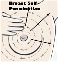
It is very important for every woman above aged 30 years old to learn, and practise BSE regularly once a month, about a week after each menstrual period. Women who no longer have periods should practise BSE on a fixed date each month.

2.Mammography
------------------
1.Women aged 40 - 49 years are encouraged to go for regular mammography every year 2.Women 50 years and above should go every two years.
3.Women who are at higher risk of developing breast cancer should see a doctor for advice. You may need to go for screening earlier and more frequently.
Mammography is a low-dose X-ray of the breast using specially designed X-ray machine.
It is the most effective method to detect very small lumps in the breast even before they can be felt by the hand.

3.Ultrasound of the Breasts
-------------------------------
Ultrasound of the Breasts is used together with mammography in cases where the diagnosis of possible cancer of the breast is not confirmed.
The ultrasound may be able to detect small lumps, which are not detected by mammogram.

4.MRI (magnetic resonance imaging) of the Breasts
----------------------------------------------------------
MRI is a procedure that uses a magnet, radio waves, and a computer to make a series of detailed pictures of areas in the body. With advancement of technology, a MRI of the breasts can detect even more clearly any abnormal lumps or tissues in the breasts.

5.Estrogen and progesterone receptor test:
-----------------------------------------------
A test to measure the amount of estrogen and progesterone (female hormones) receptors in cancer tissue. The test results show whether hormone therapy may stop the cancer from growing.

6.Biopsy:
-----------
The removal of cells or tissues so they can be viewed under a microscope by a pathologist to check for signs of cancer. If a lump in the breast is found, the doctor may need to cut out a small piece of the lump.
Four types of biopsies are as follows:
Excisional biopsy: The removal of an entire lump or suspicious tissue.
Incisional biopsy: The removal of part of a lump or suspicious tissue.
Core biopsy: The removal of part of a lump or suspicious tissue using a wide needle.
Needle biopsy: The removal of part of a lump, suspicious tissue, or fluid, using a thin needle.

What is the Treatment of Breast Cancer?
-----------------------------------------------

There are different types of treatment for patients with breast cancer.
Four types of standard treatment are used:
1.Surgery
---------
Most patients with breast cancer have surgery to remove the cancer from the breast. Some of the lymph nodes under the arm are usually taken out and looked at under a microscope to see if they contain cancer cells.
Lumpectomy: Breast-conserving surgery to remove a tumor (lump) and a small amount of normal tissue around it.
Partial mastectomy: Surgery to remove the part of the breast that has cancer and some normal tissue around it. This procedure is also called a segmental mastectomy.
Total mastectomy: Surgery to remove the whole breast that has cancer. This procedure is also called a simple mastectomy. Some of the lymph nodes under the arm may be removed for biopsy at the same time as the breast surgery or after. This is done through a separate incision.
Modified radical mastectomy: Surgery to remove the whole breast that has cancer, many of the lymph nodes under the arm, the lining over the chest muscles, and sometimes, part of the chest wall muscles.
Radical mastectomy: Surgery to remove the breast that has cancer, chest wall muscles under the breast, and all of the lymph nodes under the arm. This procedure is sometimes called a Halsted radical mastectomy.
Even if the doctor removes all the cancer that can be seen at the time of the surgery, some patients may be given radiation therapy, chemotherapy, or hormone therapy after surgery to kill any cancer cells that are left. Treatment given after the surgery, to increase the chances of a cure, is called adjuvant therapy.
If a patient is going to have a mastectomy, breast reconstruction (surgery to rebuild a breast's shape after a mastectomy) may be considered. Breast reconstruction may be done at the time of the mastectomy or at a future time.
2.Radiation therapy
--------=---------------
Radiation therapy is a cancer treatment that uses high-energy x-rays or other types of radiation to kill cancer cells or keep them from growing.
External radiation therapy uses a machine outside the body to send radiation toward the cancer.
Internal radiation therapy uses a radioactive substance sealed in needles, seeds, wires, or catheters that are placed directly into or near the cancer.
3.Chemotherapy
--------------------
Chemotherapy is a cancer treatment that uses drugs to stop the growth of cancer cells, either by killing the cells or by stopping them from dividing.
4.Hormone therapy
-----------------------
Hormone therapy is a cancer treatment that removes female hormones or blocks their action and stops cancer cells from growing.
Examples are tamoxifen and aromaase inhibitors.

Other newer methods are:
Sentinel lymph node biopsy followed by surgery
--------------------------------------------------------
Sentinel lymph node biopsy is the removal of the sentinel lymph node during surgery. The sentinel lymph node is the first lymph node to receive lymphatic drainage from a tumor. After that biopsy, the surgeon removes the tumor (breast-conserving surgery or mastectomy).
Monoclonal antibodies as adjuvant therapy
---------------------------------------------------
Monoclonal antibody therapy is a cancer treatment that uses antibodies made in the laboratory, from a single type of immune system cell. The antibodies attach to the cancer cells, block their growth, or keep them from spreading.
Examples are Trastuzumab (Herceptin) which blocks the effects of the growth factor protein HER2, Tyrosine kinase inhibitors which block signals needed for tumors to grow, Lapatinib which blocks the effects of the HER2 protein and other proteins inside tumor cells.

Treatment of Preinvasive Breast Cancer
------------------------------------------------

Surgery is the main form of treatment for this stage of Cancer.
Either a lumpectomy or a mastectomy can be done.
Women who choose lumpectomy usually have to undergo radiation treatment or radiotherapy.

The type of surgical treatment depends on
1. The size and extent of cancer within the breast
2.the patient's suitability or desire to undergo radiotherapy.

Chemotherapy
is not required for cancer detected at such an early stage.

Treatment of Invasive Breast Cancer
-------------------------------------------

Surgery is the main treatment as well in this case.
It involves the removal of the cancer with or without preserving the breast.
In the late stages where the cancer has spread to the other parts of the body, surgical removal of the cancer may be done to reduce the pain.

Chemotherapy is given to treat the cancer that has spread beyond the breast and armpit. It can be given orally or by injection.
The decision to use chemotherapy depends on:
1. The stage of the cancer
2. Type of cancer cells
3. Whether the woman has reached menopause
Chemotherapy may be used before surgery in large cancers to reduce cancer recurrence or to increase the chance of safe breast preservation surgery.

Hormonal therapy is used for hormone responsive cancers to deprive the cancer cells of oestrogen, which is required for such cancers to grow.
It is given in place of chemotherapy or following it.

Radiotherapy uses high-energy rays to kill cancer cells or stop them from growing further. It may be given for a peroid of time after a lumpectomy or after a mastectomy.
It is also given for inoperable cancers.

How is Breast Cancer Recurrence detected?
---------------------------------------------------
After cancer treatment, patients are required to follow up with their doctors at regular intervals especially in the first 5 years after treatment when the risk of recurrence is highest.
This include:
Physical examination of the breasts, underarm, neck.
Peroidic mammograms
Blood tests
Chest x-rays and
Bone scan.
Any complaints of change in surgical areas or other parts of the breasts, swollen lymph glands or bone pains may suggest a recurrence.

What is the Prognosis of Breast Cancer?
-----------------------------------------------
The prognosis (chance of recovery) and treatment options depend on the following:
1.stage of the cancer (whether it is in the breast only or has spread to lymph nodes or other places in the body).
2. type of breast cancer.
3.Estrogen-receptor and progesterone-receptor levels in the tumor tissue.
4.woman's age, general health, and menopausal status (whether a woman is still having menstrual periods).
5.Whether the cancer has just been diagnosed or has recurred.

Finally please note that Breast Cancer can also occur in Men!
Thursday, August 30, 2007

A Simple Guide to Psoriasis


A Simple Guide to Psoriasis
---------------------------------


What is Psoriasis?
---------------------

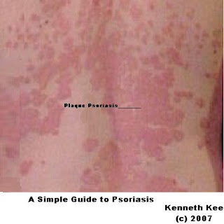
Psoriasis is a disorder of the skin which typically consists of red patches covered by silvery-white scales especially around the trunk, elbows, knees and scalp.

What are the types of Psoriasis?
--------------------------------------

There are five types, each with unique signs and symptoms.

1.Plaque psoriasis is the most common type of psoriasis.
About 80% of people who develop psoriasis have plaque psoriasis, which appears as patches of raised, reddish skin covered by silvery-white scale.
These patches, or plaques, frequently form on the elbows, knees, lower back,and scalp.

2.guttate psoriasis (small, red spots on the skin),

3.pustular psoriasis (white pustules surrounded by red skin),

4.inverse psoriasis (smooth, red lesions form in skin folds)

5.erythrodermic psoriasis (widespread redness, severe itching, and pain).

Who Gets Psoriasis?
-----------------------

Psoriasis occurs about equally in males and females.
Recent studies show that there may be an ethnic link.
It seems that psoriasis is most common in Caucasians and slightly less common in African Americans.
Worldwide, psoriasis is most common in Scandinavia and other parts of northern Europe.
It appears to be far less common among Asians and is rare in Native Americans.

There also is a genetic component associated with psoriasis.
About one-third of people who develop psoriasis have at least one family member with the condition.

Research shows that the signs and symptoms of psoriasis usually appear between 15 and 35 years of age.
About 75% develop psoriasis before age 40.
However, it is possible to develop psoriasis at any age. After age 40, a peak onset period occurs between 50 and 60 years of age.

About 1 in 10 people develop psoriasis during childhood, and psoriasis can begin in infancy.
The earlier the psoriasis appears, the more likely it is to be widespread and recurrent.

What causes Psoriasis?
---------------------------

Its exact cause is unknown but the result is skin which grows about seven times more quickly and thicker than usual.
It is thought to be due, at least in part, to an abnormal immune reaction against some component of the skin.
Genetic factors play a part.
About half those affected know of someone else in the family with it.

The rash often seems to start after some sort of trigger. This may be emotional stress, skin injury (cuts and scratches for example), a streptococcal sore throat, hormones (it often occurs at puberty), or rarely, certain medications.
Psoriasis is not contagious.

What are the symptoms of Psoriasis?
--------------------------------------------

Itchy, raised patches of dark pink skin.
White scales on the scalp, knees, elbows and upper body.
Pitting, discolouring and thickening of the fingernails and toenails.
Red, scaly and cracked skin on the palms of the hands or on the soles of the feet.
There may be fluid oozing out of the cracks and movement may become very painful. The itching and discomfort may keep a person awake at night, and the pain can make everyday tasks difficult.

How does Psoriasis affects the Patient?
-----------------------------------------

All types of psoriasis, ranging from mild to severe, can affect a person’s quality of life.
Living with this lifelong condition can be physically and emotionally challenging.
Itching, soreness, and cracked and bleeding skin are common.
Nail psoriasis can be painful.
Even the simple act of squeezing a tube of toothpaste can hurt.
Several studies have shown that people often feel frustrated.
In some cases, psoriasis limits activities and makes it difficult to perform job responsibilities.
Studies also have shown that stress, anxiety, loneliness, and low self-esteem are part of daily life for people living with psoriasis.
It has been that thoughts of suicide are three times higher for psoriatics than the normal person.
Embarrassment is another common feeling.
What if you extended your hand to someone and the person recoiled?
How would you feel if you spent most of your life trying to hide your skin?

What is the Treatment of Psoriasis?
------------------------------------------

Psoriasis is a chronic, meaning lifelong, condition because there is currently no cure. People often experience flares and remissions throughout their life.
Controlling the signs and symptoms typically requires lifelong therapy.
Treatment depends on the severity and type of psoriasis.
Some psoriasis is so mild that the person is unaware of the condition.
A few develop such severe psoriasis that lesions cover most of the body and hospitalization is required. These represent the extremes. Most cases of psoriasis fall somewhere in between.

Topical medications (such as steroid and tar lotions, ointments, creams, and shampoo)
Photo therapy (either ultraviolet B, or UV, radiation or psoralen with ultraviolet A, or PU VA, radiation)
Systemic therapy (oral drugs that affect the whole body system, not just the skin e.g. cyclosporins, methotrexate)

Combinations are often used. These treatments are often given in the order described -- from least to most potent -- as necessary, although the physician may choose a more powerful therapy or use combinations first if the patient's initial condition is severe.

With the emergence of several new therapies, including the biologic agents, more people are experiencing substantial improvements and reporting a greatly improved quality of life.

A Simple Guide to Eczema


A Simple Guide to Eczema
-------------------------------


What is Eczema?
--------------------

Eczema is a general term meaning inflammation of the skin with signs of redness, itch and swelling.
One of the most common forms of eczema is atopic eczema which affects 10-20% of the world population. It occurs at some point during childhood.
Fortunately, many children with eczema find that the disease clears and often disappears with age.

What causes Eczema?
--------------------------


There is no known cause for the disease.
It often affects people with a family history of allergies.
Many people who have eczema also suffer from allergic rhinitis and asthma, or have family members who do.
Those who are genetically predisposed may deverlop eczema when exposed to environmental and food triggers:
milk,egg white,nuts, shellfish
dust, chemicals,detergents,flowers,pollens,dyes in clothes,cosmetics

Who gets eczema?
----------------------


Eczema occurs in both children and adults, but usually appears during infancy.

Is Eczema contagious?
---------------------------


Eczema is not contagious and, like many diseases, currently cannot be cured.
For most patients the condition may be managed well with treatment and avoidance of triggers.

How common is eczema?
------------------------------


The condition is quite common.
About10-20% of all infants have eczema.
In nearly half of these children, the disease will improve greatly by the time they are between five and 15 years of age.
Others will have some form of the disease throughout their lives.

Where does Eczema appear?
-----------------------------------

Eczema can occur on just about any part of the body.
In infants, eczema typically occurs on the forehead, cheeks, forearms, legs, scalp, and neck.
In children and adults, eczema typically occurs on the face, neck, and the insides of the elbows, knees, and ankles.
In some people, eczema may form bubbles which breaks and ooze.
In others, the condition may appear more scaly, dry, and red.
Chronic scratching causes the skin to take on a leathery texture because the skin thickens (lichenification).

What are the symptoms and signs of eczema?
-----------------------------------------------------


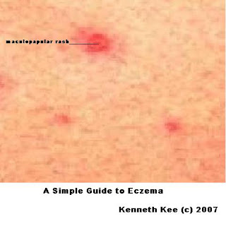
Typically there are dry, red, extremely itchy patches on the skin.
Eczema is sometimes referred to as "the itch that rashes," since the itch, when scratched, results in the appearance of the rash.

What makes patients with eczema itch?
-----------------------------------------------

The triggers that causes a flare up of the eczema causes irritation to the skin.
When the skin becomes irritated, histamines are released into the skin causing the redness and itchiness.
For some, rough or coarse materials coming into contact with the skin causes itchiness. For others, feeling too hot and sweating will cause an outbreak.
Other people find that certain soaps, detergents, disinfectants, contact with juices from fresh fruits and meats, dust mites, and animal furs may trigger itching.
Upper respiratory infections (caused by viruses) may also be triggers.
Stress can also sometimes aggravate an existing flare-up,a condition called neurodematitis.

How can eczema be prevented?
-------------------------------------


Eczema outbreaks can usually be prevented with some simple precautions:
1.Moisturize frequently
2.Avoid sudden changes in temperature or humidity
3.Avoid sweating or overheating
4.Avoid scratchy materials (e.g., wool or other irritants)
5.Avoid harsh soaps, detergents, and solvents
6.Avoid environmental factors that trigger allergies (e.g., pollens, molds, mites, and animal furs)
7.Be aware of any foods that may trigger an attack and avoid those foods
8.Reduce stress

How can eczema be treated?
----------------------------------


The most important part of an eczema treatment routine is to avoid scratching because that will irritate the skin even more.

The most common treatment is the application of lotions or creams to keep the skin as moist as possible. These treatments are generally most effective when applied directly after bathing so that the moisture from the bath is remains in the skin.

Cold compresses applied directly to itchy skin can also help relieve itching.

Another effective treatment is the application of corticosteroid creams and ointments to reduce inflammation.To prevent side effects such as skin thinning, your doctor may limit the length of treatment time and locations where you can apply treatment.

For severe eczema, your doctor may prescribe oral corticosteroids, but be aware that side effects including new flare-ups can develop when treatment is discontinued (this treatment is not recommended for long-term use).

Skin affected by eczema may frequently become infected. A course of topical or oral antibiotics may be prescribed to kill the bacteria causing the infection.

For severe itching, sedative and non-sedative antihistamines are sometimes used to reduce the itch.
For day time activities as well as driving, non sedative antihistamine should be used.
Because drowsiness is a common side effect, sedative antihistamines are often used in the evening to help a person restless from eczema get to sleep.

Tar treatments and phototherapy are also used and can have positive effects. Phototherapy requires special equipment (lights).

Finally, in cases where eczema is resistant to therapy, your physician may prescribe the drug cyclosporine A, which modifies immune response.
This is used only in extreme cases because of its association with serious side effects.

What can be done for children with eczema?
----------------------------------------------------


Children are special because it is difficult for them to resist scratching their eczema.
For mild to moderate cases, the application of moisturizer on a regular basis can be very helpful. And, in most cases, the eczema will disappear as the child ages.
In the meantime, avoid as many eczema triggers as possible.
Keep your child’s skin moist. After bathing, apply moisturizer to retain the moisture in the skin.
Avoid sudden temperature changes.
Keep your child’s bedroom and play areas free of dust mites.
Use mild soaps – both on your child’s skin and on your child’s clothing.
Dress your child in breathable, preferably cotton, clothing.

Tuesday, August 28, 2007

A Simple Guide to Vasomotor rhinitis


A Simple Guide to Vasomotor rhinitis
--------------------------------------

What is Vasomotor Rhinitis?
-----------------------------


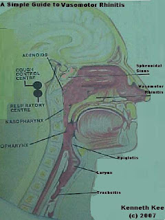
Vasomotor Rhinitis is a condition which consists of a group of symptoms that include a runny nose, itchiness and sneezing, that are caused by irritation and congestion in the nose. It is brought about by changes in vascular tone and permeability.

What are the symptoms of Vasomotor rhinitis?
--------------------------------------------------

The symptoms of Vasomotor rhinitis include:
Recurring nasal inflammation
Sneezing
Runny nose
Profuse watery nasal discharge
Nasal membrane swelling

What causes Vasomotor rhinitis?
---------------------------------------

Vasomotor rhinitis occurs as a result of a response of the nasal membrane to
1.irritants such as smoke,
2.temperature changes and
3.stress.

Other causes of chronic rhinitis are blockage in the nose e.g. nasal polyps, enlarged adenoids and a deviated septum that impede mucus drainage and restrict air flow. Medications can cause rhinitis or worsen it in people with allergies, vasomotor rhinitis or a deviated septum.
Prolonged use of nasal decongestants can worsen rhinitis.
Some blood pressure medications such as beta blockers and vasodilators can cause rhinitis.

What is the difference between Allergic rhinitis and Vasomotor Rhinitis?
--------------------------------------------------------------------------------------

Allergic rhinitis could be seasonal or perennial (year round).
Seasonal rhinitis is due to pollen or mould spores.
Perennial rhinitis are due to allergens such as house dust mites, animal dander or moulds found on carpets or furniture upholstery and even some foods.

Non-allergic rhinitis can also be triggered by cigarette smoke and many other air pollutants, strong odours, exposure to cold and alcohol. Smoking and air pollution may also cause symptoms by damaging the cilia (hair cells) which moves mucus through the air passages.

Hormonal changes in pregnancy cause "Rhinitis in Pregnancy". The expecting mother will recover once the baby is delivered.

What treatments are available for Vasomotor rhinitis?
----------------------------------------------------------------

Treatments for Vasomotor rhinitis include:
Non-medical:
----------------
Avoid dry air
Keeping air moist
Vapourisers
Nasal Decongestant sprays are not recommended for treatment of vasomotor rhinitis

Complete avoidance of environmental allergens may be impossible but it can be minimised for example removing carpets and pets.
Use a pollen mask when mowing the grass or cleaning the house
Install an air purifier
Change the air filters monthly in heating and air conditioning systems
use cotton or synthetic materials such as Dacron in pillows and bedding
enclose mattress in plastic
keep windows closed during high pollen times
eliminate house plants
bathe pets frequently or even give away dander–producing pets.

Avoid Nasal irritants which can cause typical immune response seen with classical allergies. Examples include cigarette smoke, perfume, aerosol sprays, smoke, and smog and car exhaust

Nasal irrigation utilizing a buffered hyper tonic saline solution helps to reduce swollen and congested nasal and sinus tissues.
It also washes out thickened nasal secretions, irritants (smog, pollens, etc.), bacteria, and crusts from the nose and sinuses.
While irrigating the nose, it is best to stand over the sink and irrigate each side of your nose.
Aim the stream toward the back of your head, not at the top of your head.
For young children, the salt water can be put into a small spray container which can be squirted many times into each side of the nose.

Medical:
---------

Anti-histamines provide good relief for mild to moderate symptoms.
Newer antihistamines that are long-acting and less likely to cause drowsiness have been found to be useful.

Decongestants such as pseudoephedrine can help to shrink the swollen tissues caused by irritants and other causes. However decongestant nasal sprays are not advised because they cause a a rebound effect on nasal tissues(initially the nasal tissue shrinks but later they become more swollen)

Corticosteroids in the form of nasal spray reduce the immune response and may be prescribed to reduce severe symptoms.
These drugs are highly effective in allergic patients; but there is a potential for serious side effects when used over time. They are best used for the short term management of allergic problems, and their use must always be monitored by a physician.

Nasalcrom (cromolyn sodium): This spray helps to stabilize allergy cells (mast cells) by preventing release of allergy mediators, like histamine.

Immunotherapy:Allergy shots interfere with the allergic response. After identification of an allergen, small amounts of it are given back to the sensitive patient. Over time the patient will develop blocking antibodies to the allergen, and they become less sensitive and less reactive to the substance causing allergic symptoms. This is the best treatment provided the allergen is correctly identified.

Surgery:Nasal polyps, enlarged adenoids and deviated septums can be corrected with surgery. Obviously this should be done only after more conservative measures have been tried. Surgery is not a replacement for good allergy control and treatment.

A Simple Guide to Anaemia


A Simple Guide to Anaemia
--------------------------------


What is Anaemia?
----------------------


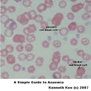
Anaemia is a blood condition in which the haemoglobin is lower than the average value of a normal person. The Haemoglobin is the red chemical in red blood cell which carries oxygen around our bodies.

What Causes Anaemia?
----------------------------


The commonest causes of anaemia is:

1. blood loss such as excessive menstrual flow or
internal bleeding from the stomach or bowel

2. deficiency in the diet for foods rich in iron, vitamin B12 or folic acid.

3.inherited abnormalities (Thalassaemia)

4. kidney failure ,liver disease

5. cancer patients

5.certain drugs such as anti cancer drugs and toxic substances such as lead

6. infections (haemorhagic dengue fever) & worm infestation

7.radiation therapy,

8.surgery on the gastro-intestinal tract, particularly the stomach.

What are Symptoms of Anaemia?
----------------------------------------


Easy fatigue :
This is due to lack of oxygen in the red blood cells
Pale skin :
The lack of haemoglobin cause the redness of the blood cells to be reduced hence the pale appearance of the skin.The finger nails and lower eyelids can be checked for the paleness.
Dizziness:
Giddiness occurs when there is insufficient oxygen supply to the nerve cells in the brain.
Unusually rapid heart beat:
Rapid heart beat may occur becuse the heart has to pump faster to supply enough oxygen to the body
Difficulty concentrating and headache:
These are caused by insufficient oxygen to the brain.
Leg cramps
Leg cramps are due to insufficient oxygen to the muscles.
Shortness of breath:
Breathing difficulty may occur as a result of insufficient oxygen in the lungs

What is the Treatment of Anaemia?
------------------------------------------


1.Treat the underlying cause of the anaemia.

2.Eat a well-balanced diet that includes good sources of
iron (liver, meat, peas, beans, whole grain),
vitamin B12 (foods of animal origin only, including meats, liver and kidney, milk, cheese, fish, shell fish and eggs)
folic acid( present in most vegetables).
Vitamin C can make the stomach more acidic and can improve the absorption of iron in your diet.

3.Reduce your consumption of caffeine products and tea as they can decrease the absorption of iron.

Saturday, August 25, 2007

A Simple Guide to Uveitis


A Simple Guide to Uveitis
-------------------------------

What is Uveitis?
------------------

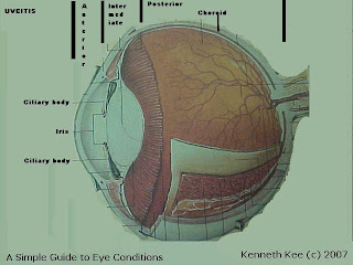
Uveitis is inflammation of the eye's uvea. The uvea is the eye's middle layer that consists of the iris, ciliary body and choroid.

What are different types of Uveitis?
----------------------------------

Uveitis is classified by the part of the uvea it affects.

1. Anterior uveitis refers to inflammation of the iris alone (called iritis) or the iris and ciliary body. Anterior uveitis is the most common form.

2.Intermediate uveitis refers to inflammation of the ciliary body.

3. Posterior uveitis is inflammation of the choroid.

Diffuse uveitis is inflammation in all areas of the uvea.

What are the dangers of Uveitis?
--------------------------------------

Many cases of uveitis are chronic.
They can produce numerous possible complications, including clouding of the lens (cataract) or cornea, elevated intraocular pressure (IOP), glaucoma, and retinal problems (such as swelling of the retina or retinal detachment).
These complications can lead to loss of vision.

Who are affected by Uveitis?
----------------------------------

Uveitis occurs most frequently in people ages 20 to 50.
It is more common in women and more likely to develop with age.

What are the Complications of Uveitis?
----------------------------------------------

Uveitis complications:
secondary cataracts,
secondary glaucoma,
high intraocular pressure,
retinal detachment

What are the Symptoms and Signs of Uveitis?
-----------------------------------------------------

With anterior uveitis, the symptoms usually affect only one eye.
There may be
mild to strong pain
redness and
light sensitivity.
Your vision may also be blurred.
Eye is tender to palpation

Both intermediate and posterior uveitis are usually painless.
Symptoms are blurred vision and floaters, typically in both eyes.

Diffuse uveitis has a combination of symptoms of all types of uveitis.

What Causes Uveitis?
--------------------------

Uveitis has dozens of causes, including viral, fungal, and bacterial infections.
But, in many cases, the cause is unknown (idiopathic).
Eye doctors can sometimes identify the cause if there has been trauma to the eye, such as from surgery or a blow, or if there is a history of an infectious or immunological systemic disorder.
Some of the many different systemic disorders that can cause uveitis include:
acute posterior multifocal placoid pigment epitheliopathy
ankylosing spondylitis
Behçet's disease
birdshot retinochoroidopathy
brucellosis
herpes simplex
herpes zoster
inflammatory bowel disease
juvenile rheumatoid arthritis
Kawasaki's disease
leptospirosis
Lyme disease
multiple sclerosis
presumed ocular histoplasmosis syndrome
psoriatic arthritis
Reiter's syndrome
sarcoidosis
syphilis
systemic lupus erythematosus
toxocariasis
toxoplasmosis
tuberculosis
Vogt-Koyanagi-Harada syndrome

What is the Treatment of Uveitis?
---------------------------------------
Non-drug:
-------------
warm water
pads to cover eyes for rest and avoid sun

Drugs:
--------

Your doctor will likely prescribe a steroid to reduce the inflammation in your eye.
Whether the steroid is in eye drop, pill, or injection form depends on the type of uveitis you have.
Because anterior uveitis affects the front of the eye, it's easy to treat with eye drops.
Intermediate uveitis can go either way, and
posterior uveitis usually requires tablets or injections.

Steroids and other immunosuppressants can produce many serious side effects:
such as
kidney damage,
high blood sugar,
high blood pressure,
osteoporosis, and
glaucoma.

In April 2005 the drug Retisert was approved by FDA for chronic non-infectious posterior uveitis. Retisert is a drug implant and is the first of its kind for treatment of uveitis. It consists of a drug reservoir that delivers sustained amounts of fluocinolone acetonide, an anti-inflammatory corticosteroid, for about two and a half years. It is implanted into the back of the eye. During clinical trials the incidence of reccurent uveitis fell from 40%-54% to 7%-14%. The most common side effects noted during those studies were cataract progression, increased intraocular pressure, procedural complications, and eye pain.

If you have anterior uveitis, your doctor will likely prescribe, in addition to the steroids, pupil-dilating eye drops to reduce pain.
You may also need eye drops to lower your intraocular pressure if it's elevated.

Friday, August 24, 2007

A Simple Guide to Parkinson's Disease


A Simple Guide to Parkinson's Disease
----------------------------------

What is Parkinson's Disease?
----------------------------------
Parkinson's Disease is a disorder of old age characterised by slow movement , rest tremors , rigidity and poor coordination.

Who gets Parkinson's Disease?
------------------------------------

Parkinson's Disease usually after the age of 50 years.
Incidence is about 0.2% of population.
It is one of the most common neurologic disorders of the elderly.
It affects both men and women equally.

What causes Parkinson's Disease?
---------------------------------------

Parkinson's Disease results when the nerve cells in the part of the brain that controls muscle movement (substantia nigra) are gradually destroyed.
Nerve cells use a brain chemical called dopamine to help send signals back and forth.
Damage in the area of the brain that controls muscle movement causes a decrease in dopamine production.
Low dopamine affects the balance between nerve-signalling substances (transmitters).
As a result, the nerve cells cannot properly send messages.
This results in the loss of muscle function.

What are the symptoms of Parkinson's Disease?

--------------------------------------------------------

Early symptoms may be nonspecific and may include numbness, painful and tender muscles, stiffness and weakness of limbs, fatigue and unexplained weight loss.

As the disease progresses, the classical features of Parkinson's Disease appear:
1. Bradykinesia(slowness of movement)
2. leadpipe rigidity
3. rest tremors
4. Postural instability


How is Parkinson's Disease diagnosed?
----------------------------------------------

Parkinson's Disease is diagnosed when there are at least 2 of the four classical features are present. To improve diagnostic accuracy, bradykinesia must be present.

1. Bradykinesia usually presents with lack of voluntary and automatic movement such as monotonous speech,
lack of facial expression,
Slow movements
Difficulty initiating any voluntary movement
Difficulty beginning to walk
Difficulty getting up from a chair
shuffling of feet movement

One way to demonstrate this is to have the patient tap his index finger on his thumb.
The movement is slow and lack momentum.

2. Rigidity of Parkinson's Disease is leadpipe in nature.
This rigidity contribute to the
mask like facial appearance,
Muscle rigidity
stiffness of posterior neck muscles,
diminished arm swing,
cogwheel rigidity of the wrists,
Difficulty bending arms or legs

3.Rest tremor is classical of Parkinson's Disease.
There is this resting or pill rolling tremors of the hand which is worsened by anxiety and fatigue and disappears when the hand is in use. Tremor is usually unilateral.

4. Postural instability usually occurs late in the illness.
It is seen in the
Unstable, stooped, or slumped-over posture
freezing gait,
retropulsion,
loss of balance and
frequent falls

Turning in bed, rising from the chair and turning when walking is difficult because of poor body and limbs coordination.

What are the other symptoms associated with Parkinson's Disease?
-----------------------------------------------------------------------------

Additional symptoms that may be associated with this disease:
Depression
Confusion
Dementia
Seborrhea (oily skin)
Loss of muscle function or feeling
Muscle atrophy
Memory loss
Drooling
Anxiety, stress, and tension

What are the Complications of Parkinson's Disease?

------------------------------------------------------------

Untreated Parkinson's Disease progresses to total disability, often accompanied by general deterioration of all brain functions, and may lead to an early death.

Treated, the disorder impairs people in varying ways.
Most people respond to some extent to medications.
The side effects of medications may be severe:
Varying degrees of disability
Difficulty swallowing or eating
Difficulty performing daily activities
Injuries from falls
A variety of gastrointestinal symptoms, mainly constipation
Daily activities such as bathing,dressing, walking and even writing may be difficult.

How is Parkinson's Disease treated?
------------------------------------------

Parkinson's Disease is not curable and the purpose of treatment is to:
1. improve functions and
2. treat symptoms

Treatment of Parkinson's Disease can be broadly classified into:
1. Non-drug therapy:

Good general nutrition and health
are important.
Exercises that improve strength, agility and flexibility are useful to minimise the disability of Parkinson's Disease.
A range of motion exercises is useful to keep the joints supple and mobile.
Patients are encouraged to exercise according to their ability.
Good exercises include walking,swimming, stretching and riding stationary bicycles.

Patients are taught sitting balance,walking techniques and the use of handrails. Activities of daily living is made more manageable with aids like:
Buttoning -replace buttons with Velcros
Eating -use utensils with large handles
Getting out of bed -install an overhead grab bar
Prevents falling -install hand bars in bedroom and bathroom. use nonslip mats.
Getting out of chair - use high seat chair
Railings or banisters placed in commonly used areas of the house may be of great benefit to the person experiencing difficulties with daily living activities.
Special eating utensils may also be helpful.

2. Drug therapy

There are 2 main types of drugs:
1. symptom modifying helps to alleviate the symptoms but do not change the natural history of the conditions.
depression
insomnia
constipation,
dysphagia,
urinary frequency

2. disease modifying drugs such as:
Levodopa may be used to increase the body's supply of dopamine, which may improve movement and balance.
Artane -is particularly good for reducing tremors
Jumex- is used in early Parkinson's Disease and helps reduce bradykinesia
Bromocriptine - enhance the effect of levadopa Deprenyl may provide some improvement to mildly affected patients.
Amantadine or anticholinergic medications may be used to reduce early or mild tremors. Carbidopa reduces the side effects of levodopa and makes levodopa work better.
Entacapone is used to prevent the breakdown of levodopa.
Pramipexole and ropinirole are used before or together with levodopa.
Rasagiline is approved for patients with early Parkinson's disease. Rasagiline helps block the breakdown of dopamine.
Neupro is a new skin patch that contains the drug rotigotine. This medicine helps dopamine receptors in the brain work better. The patch is replaced every 24 hours.

Additional medications to help reduce symptoms or control side effects of primary treatment medications include antihistamines, antidepressants, monoamine oxidase inhibitors (MAOIs), and others.
It is important that the disease should be controlled before irreversible brain damage occurs.

3. Surgery:
In some cases surgery can help to alleviate symptoms especially when all other medications and measures failed.
Surgery to implant stimulators or destroy tremor-causing tissues may reduce symptoms in some people.
Transplantation of adrenal gland tissue and stem cells to the brain has been attempted, with variable results.


Added 4th October 2008:
---------------------------------

Parkinson Disease:
----------------------
Less than 5% of Parkinson's Disease patients gets the disease through genetic reasons.

A mutation of a gene (LRRK2) called G20135 has been found to increase the risk of Parkinson's disease in some families.

People with the mutated gene gets the illness at a younger age than the normal which is 60 and above.

The cause of Parkinson's disease is still unknown.

The prevailing theory is that environmental factors such as exposure to high levels of pesticides is the cause of the disease.

Thursday, August 23, 2007

A Simple Guide to Stye & Chalazion


A Simple Guide to Stye & Chalazion
------------------------------------

What is a Stye?
----------------
A Stye is an infected gland at the edge of the eyelid.

How does a Stye develop?
-----------------------------

A stye develops when a gland supplying wax to eyelash at the edge of the eyelid becomes blocked and infected.
A stye can grow on the inside or outside of the eyelid.
Styes are not harmful to vision.
They can occur at any age.

What are the Symptoms and Signs of Stye?
--------------------------------------------------

A stye starts with pain, redness, tenderness and swelling in the area of the oil gland of the eyelid. Then a small pimple appears. Sometimes just the immediate area is swollen.
At other times the entire eyelid swells.
There may be frequent tearing in the affected eye, a feeling like something is in the eye or increased light sensitivity.

What Causes Styes?
-----------------------

Most Styes are caused by staphylococcal bacteria.
This bacterium is often found in the nose, and it's easily transferred to the eye by rubbing first your nose, then your eye.
Styes are not normally harmful to vision
They generally heal within a few days.

What is the Treatment for Stye?
--------------------------------------

Most styes heal within a few days on their own.
You can apply hot compresses for 10 to 15 minutes, three or four times a day over the course of several days. This will relieve the pain and bring the stye to a head, much like a pimple. The stye ruptures and drains, then heals.

If you have frequent styes, your eye doctor may prescribe a course of antibiotic and ask you to apply an antibiotic eye ointment to kill the bacteria causing the stye.

In most cases, the Styes formed inside the eyelid either disappear completely or rupture on their own. In some cases, the styes become bigger and can be more serious.
These styes may need to be surgically incised and drained by your eye care practitioner.

What is a Chalazion?
-------------------------

Chalazion is an another type of Eyelid swelling usually inner to the edge of the eyelid.
Often mistaken for a stye, a chalazion is an enlarged, blocked oil gland (tarsal gland)in the inner lining of the eyelid and not at the edge of the eyelid as in the case of a stye.

A chalazion may look like a stye for the first few days, then turns into a hard painless lump in the inner eyelid later on.
Most chalazion develops further from the eyelid edge than stye.

The same treatment is used for the healing of a chalazion.
However in this case, the swelling may linger for one to several months.
If the chalazion remains after several months, your eye doctor may surgically incise and drain it to facilitate healing.

Wednesday, August 22, 2007

A Simple Guide to Rheumatoid Arthritis


A Simple Guide to Rheumatoid Arthritis
-----------

What is Rheumatoid Arthritis?
-----------

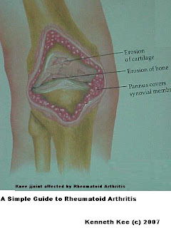
Rheumatoid arthritis is a systemic autoimmune disease that can cause chronic inflammation of the joints and other areas of the body.

Who gets Rheumatoid Arthritis?
--------------------------------------

Rheumatoid Arthritis is one of the most common form of inflammatory arthritis.
It most commonly occurs between the 25 and 50 years.
However it can occur at any age.
Females are 3 times more likely to get Rheumatoid Arthritis than males.

What causes Rheumatoid Arthritis?
-------------------------------------------

Rheumatoid Arthritis is an autoimmune disease..
Autoimmune diseases are illnesses that occur when the body tissues are mistakenly attacked by its own immune system which consists of cells and antibodies designed normally to "seek and destroy" invaders of the body, particularly infections.
Patients with autoimmune diseases have antibodies in their blood that target their own body tissues .

What are the symptoms of Rheumatoid Arthritis?
-----------------------------------------------------------

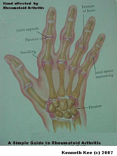
Most common symptoms are pain, swelling, stiffness, warmth and sometimes redness of the joints.

Rheumatoid Arthritis is suspected when there are:
1. Swelling in the joints especially those of the hands,elbows,knees,ankles and feet.

2. Stiffness in the joints in the mornings which can last more than an hour or after prolonged rest

3.redness and warmth in one's joints

4.Persistent pain or tenderness in a joint for more than a month

5.Inability to move or use a joint normally

6. Unexplained weight loss and appetite, fatigue, or weakness together with joint pains

What tests are done to confirm Rheumatoid Arthritis?
----------------------------------------------------------------

Abnormal blood antibodies can be found in patients with rheumatoid arthritis.

1.A blood antibody called "rheumatoid factor" can be found in 80% of patients.

2.Another antibody called "the antinuclear antibody" (ANA) is also frequently found in patients with rheumatoid arthritis.

3.Another blood test that is used to measure the degree of inflammation present in the body is the C-reactive protein.

4.A blood test called the sedimentation rate (ESR) is a measure of how fast red blood cells fall to the bottom of a test tube. The sed rate is used as a crude measure of the inflammation of the joints.

The rheumatoid factor, ANA, sed rate, and C-reactive protein tests can also be abnormal in other systemic autoimmune and inflammatory conditions. Therefore, abnormalities in these blood tests alone are not sufficient for a firm diagnosis of rheumatoid arthritis

How does the Rheumatoid Arthritis starts?
--------------------------------------------------

Rheumatoid Arthritis may start gradually or with a sudden and severe attack.
It usually attacks many joints at one time.
Rheumatoid arthritis is a chronic disease, characterized by periods of disease flares and remissions.

Where does Rheumatoid Arthritis occur?
-------------------------------------------------

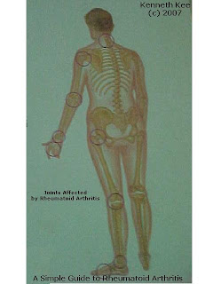
In rheumatoid arthritis, multiple joints are usually affected in a symmetrical pattern.
It most commonly affects the hands and feet,often in a systemic pattern(both right and left joints are affected).
Other joints affected are the elbow,knees,and ankles.
Less common are shoulder,hip and spine.

What are the Complications of Rheumatoid Arthritis?
----------------------------------------------------------------

Rheumatoid Arthritis can result in joint deformities and disability especially in untreated severe cases.
Daily activities such as bathing,dressing, walking and even writing may be difficult.
Besides the joints, Rheumatoid Arthritis may sometimes affect other organs such as the eyes,lungs,skin,intestines,nerves and bone marrow.
Mild fever, fatigue, loss of appetite and loss of weight may be present.
Severe Rheumatoid Arthritis is associated with an incresed risk of mortality.

How is Rheumatoid Arthritis treated?
--------------------------------------------

There is no known cure for rheumatoid arthritis.
Therefore the purpose of treatment is to:
1. improve functions and
2. reduce pain and discomfort.

Treatment of Rheumatoid Arthritis can be broadly classified into:
1. Non-drug therapy:
Weight loss is an effective way to reduce the stress on the joints and minimise the pain Walking sticks are useful ways to offload the stress on the affected weight bearing joints such as hips or knee.
Hot packs are useful aids in the morning to soften a stiff arthritic joint whereas braces and knee guards are useful supports to give some comfort to the knee.
Exercises that improve strength, agility and flexibilty are useful to minimise the disability of Rheumatoid Arthritis. A range of motion exercises is useful to keep the joints supple and mobile.
Water based exercises are a good alternative form of aerobic workout by patients afflicted by Rheumatoid Arthritis. The warm water especially is a good medium for joint mobility and together with the buoyancy of water it helps to minimse the body weight impact on the joints.
2. Drug therapy

There are 2 main types of drugs:
1. symptom modifying helps to alleviate the symptoms but do not change the natural history of the conditions. Examples are Non-steroidal Anti-inflamatory Drugs(NSAIDS) like diclofenac.

2. disease modifying drugs such as steroids, methothrexate, sulphasalazine, redaura, hydroxychloroquine have been shown to slow down the damage caused by Rheumatoid Arthritis. However they cause a lot of side effects such as anemia, abnormal white cells, kidney damage,liver damage, vision loss so the medications must be monitored all the time.
It is important that the disease should be controlled before irreversible joint damage occurs.

Newer "second-line" drugs for the treatment of rheumatoid arthritis include leflunomide (Arava), and the "biologic" medications etanercept (Enbrel), infliximab (Remicade), anakinra (Kineret), and adalimumab (Humira).

3. Surgery

Surgery is used as a last result to correct deformities and reduce disability.

The treatment of rheumatoid arthritis optimally involves a combination of patient education, rest and exercise, joint protection, medications, and occasionally surgery.

Early treatment of rheumatoid arthritis results in better outcomes.

Tuesday, August 21, 2007

A Simple Guide to Glaucoma


A Simple Guide to Glaucoma
----------------------------------

What is Glaucoma?
-----------------------

Glaucoma is a condition in which there is an increase in the pressure in the eye.

Because of the increase of this intraocular pressure, the nerve that connects the eye to the brain(the optic nerve) may be damaged.
This can result in decreased peripheral vision and, eventually, blindness.

Who is affected by Glaucoma?
----------------------------------

The following are at risk from Glaucoma:
1. age above 65 yrs
2. family history of glaucoma
3.history of serious eye injury or eye surgery
4.very short sighted
5. long term usage of steroid-containing eye drops
6. Diabetes

What is the cause of Glaucoma?
--------------------------------------

The pressure in the eye (Intraocular pressure) is increased when there is poor drainage of the fluid that flows in and out of the anterior chamber of the eye.

The higher pressure can then cause damage to the nerve.

What are symptoms of Glaucoma?
----------------------------------------

There are two major types of glaucoma:

1.Chronic Glaucoma( or Primary open angle Glaucoma )is sometimes referred to as a "silent thief of eyesight".

This is because it progress slowly and does not produce any symptom until it is very advanced. By that time irreversible damage of the optic nerve may have occured causing blurring of vision.

2.A less common form of Glaucoma known as acute closed angled Glaucoma causes sudden symptoms of intense eye pain, headaches, haloes around lights, dilated pupils, vision loss, red eyes, nausea and vomiting.

These signs may last for a few hours, then return again for another round. Each attack takes with it part of your field of vision. This requires emergency treatment.

Other variations include
3.congenital glaucoma:
It's difficult to spot signs for congenital glaucoma because the children are too young to understand. If you notice a cloudy, white, hazy, enlarged or protruding eye, consult your eye doctor
4. pigmentary glaucoma:
Pigmentary glaucoma often exhibits no symptoms at all. You may notice some pain and blurry vision after exercise. Pigmentary glaucoma affects mostly males in their mid-30s to mid-40s.
5.secondary glaucoma:
Symptoms of chronic glaucoma following an eye injury could indicate secondary glaucoma.

How is Glaucoma diagnosed?
----------------------------------


Glaucoma is diagnosed using specialised instruments.
These may show:
Raised intraocular pressure(in most cases)
Your eye pressure (intraocular pressure) will be measured with a tonometer. Some tonometers blow a puff of air onto your eye's surface.

damaged optic nerve

abnormal visual fields

How does Intraocular pressure occur in Glaucoma?
-------------------------------------------------------------

An increased IOP reading indicates a problem with the amount of aqueous humor (fluid) in the eye:
either the eye is producing too much fluid, or
it's not draining properly.

The drainage area is the angle formed between the cornea and the iris, which is why you see the word "angle" in the different glaucoma names.

Narrow-angle glaucoma occurs in less than 10% of glaucoma patients. In this form of the disease, aqueous humor cannot drain out of the eye due to very narrow drainage angles that are usually blocked by the iris. This condition can occur slowly and progressively, or very quickly.

Rapid closing of the angles, or acute angle-closure glaucoma, is a medical emergency. Optic nerve damage and vision loss will occur within hours if the angles are not opened to drain fluid and lower IOP.

Congenital glaucoma is a rare form of the disease affecting babies.
These children are born with narrow angles or some other defect in the drainage system of the eye.

Pigmentary glaucoma is caused by pigment from the iris clogging the draining angles, preventing aqueous humor from leaving the eye.

Secondary glaucoma develops after trauma to the eye that affects the drainage system. Injury, infection, inflammation, tumor or an enlarged cataract can precipitate secondary glaucoma.

What is the aim of treatment for Glaucoma?
----------------------------------------------------

Glaucoma is a lifelong disease.
Any damage to the optic that is discovered at diagnosis is irreversible.

The aim of treatment is thus to prevent further damage and vision loss.

This is done by lowering the intraocular pressure to a safe level.

How is Glaucoma treated?
-------------------------------

1. pressure lowering eye drops

You may be prescribed eye drops to lower the intraocular pressure.
Examples are prostaglandin eye drops, which will allow better flow of fluid within the eye or beta blocker eye drops which reduces the pressure in the eye.
These has to be applied diligently in order to work well.

The majority of glaucoma patients can be well controlled with one or more of these eye drops

2. laser treatment

You may need laser treatment to help lower the pressure or to manage acute angle closure Glaucoma.

3. surgery

Surgery is used only:
when eye drops and/or laser treatment fails
if you develop side effects with the eyedrops
if you need other eye surgery(eg cataract)

How is Glaucoma monitored?
-----------------------------------

You should go for regular follow up with your doctor.
He will check whether treatment is effective by:

measuring your intraocular pressure

examining the optic nerve for further damage

analysing your visual fields.
With proper monitoring and treatment most patients will be less
at risk of blindness.


Added 4th October 2008
----------------------------------

Glaucoma
-----------------
The best painkiller for glaucoma pain is Tegretol which relieve the pain in the nerves.

Acupuncture has been used to reduce the pressure in the eye as an alternative method of treatment.

Monday, August 20, 2007

A Simple Guide to Tonsillitis


A Simple Guide to Tonsillitis
----------------

What is Tonsillitis?
-----------------

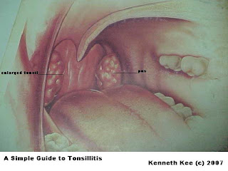
Tonsillitis is inflammation (swelling) of the tonsils.
The tonsils are lymph nodes in the back of the mouth and top of the throat.
They normally help to filter out bacteria and other germs to prevent infection in the body.
They may become so overwhelmed by bacterial or viral infection that they swell and become inflamed, causing tonsillitis.
The infection may also be present in the throat and surrounding areas, causing pharyngitis. The inflammation may involve other areas of the back of the throat including the adenoids and the lingual tonsils (areas of tonsil tissue at the back of the tongue).

What causes Tonsillitis?
-----------------------------

Viral or bacterial infections and low immunity lead to tonsillitis and its complications.
Viral:
The herpes simplex virus, Epstein-Barr virus (EBV), cytomegalovirus, adenovirus, and the measles virus cause most cases of acute pharyngitis and acute tonsillitis.
Bacteria:
Bacteria cause 15-30 percent of pharyngotonsillitis cases.
Streptococcus pyogenes is the most common bacteria causing acute Tonsillitis

Who gets tonsillitis?
------------------------

Tonsillitis most often occurs in children but rarely in children younger than 2 years.
Tonsillitis caused by Streptococcus species typically occurs in children aged 5-15 years.
Viral tonsillitis is more common in younger children.
A peritonsillar abscess is usually found in young adults but occur occasionally in children.

What are types of Tonsillitis?
----------------------------------

There are several variations of tonsillitis:
1.acute
2.recurrent
3.chronic tonsillitis and
4.peritonsillar abscess.

What are the symptoms of tonsillitis?
--------------------------------------------


The type of tonsillitis determines what symptoms will occur.

Acute tonsillitis:
Patients have a
Ear pain
Fever, chills
Headache
Sore throat - severe, lasts longer than 48 hours
Tenderness of the jaw and throat
Voice changes, loss of voice
Foul breath
dysphagia (difficulty swallowing)
Odynophagia (painful swallowing) and
tender cervical lymph nodes.

Airway obstruction due to swollen tonsils may cause mouth breathing, snoring, nocturnal breathing pauses, or sleep apnea.
Lethargy and malaise are common.

These symptoms usually resolve in three to four days but may last up to two weeks despite therapy.

Recurrent tonsillitis:
This diagnosis is made when an individual has multiple episodes of acute tonsillitis in a year.

Chronic tonsillitis:
Individuals often have
Chronic sore throat,
Foul breath,
Enlarged tonsils, and
Persistently tender cervical nodes.

Peritonsillar abscess:
Individuals often have
Severe throat pain,
fever, Drooling,
Foul breath,
Trismus (difficulty opening the mouth), and
Muffled voice quality (as if talking with a hot potato in his or her mouth).

What are the signs of Tonsillitis?
---------------------------------------

The health care provider will look in the mouth and throat for signs of:

1.enlarged, visible tonsils that are usually reddened with white spots (pus) on them.

2.enlarged and tender lymph nodes of the jaw and neck.

3.Fever and chills.

4.Open-mouth breathing and muffled voice resulting from obstructive enlarged tonsils.

5.neck and jaw stiffness (often found in acute tonsillitis).

6.Signs of dehydration (found by examination of skin and mucosa).

7.Palatal petechiae (pinpoint bleeding spots on the soft palate).

8.Unilateral bulging above and to the side of one of the tonsils in peritonsillar abscess .

A culture of the tonsils may show bacterial infection.
A culture for the streptococcus bacteria (strep) may be taken using a throat swab because it is the most common and most dangerous form of tonsillitis.

What is the treatment of Tonsillitis?
--------------------------------------------

1.If the cause of the tonsillitis is bacteria such as strep, antibiotics are given to cure the infection.
The antibiotics may need to be taken for 10 days by mouth.
They must not be stopped just because the discomfort stops, or the infection will NOT be cured.Some health care providers will treat all tonsillitis with antibiotics to prevent the chance of strep-related complications.

2.Rest to allow the body to heal.

3. Fluids especially warm (not hot), bland fluids or very cold fluids may soothe the throat. Gargle with warm salt water or suck on lozenges (containing benzocaine or similar ingredients) to reduce pain.Fluid replacement and pain control are important.

4.Hospitalization may be required in severe cases and when there is airway obstruction.

5. When the condition is chronic or recurrent, a surgical procedure to remove the tonsils (tonsillectomy) is often recommended.

What is the Prognosis of Tonsillitis?
------------------------------------------

Tonsillitis symptoms usually lessen in 2 or 3 days after treatment starts.
The infection usually is cured by then, but may require more than one course of antibiotics.
A tonsillectomy may be recommended if tonsillitis is severe, recurrent, or does not respond to antibiotics.

What are the Complications of Tonsillitis?
--------------------------------------------------

1.Complications of untreated strep tonsillitis may be severe:
Rheumatic fever and subsequent cardiovascular disorders
Post-streptococcal glomerulonephritis followed by kidney failure

2.Dehydration from difficulty swallowing fluids

3.Blocked airway from enlarged tonsils

4.Peritonsillar abscess or abscess in other parts of the throat

Sunday, August 19, 2007

A Simple Guide to Sinusitis


A Simple Guide to Sinusitis
-----------------

What is sinusitis?
----------------

Sinusitis is a condition in which the lining of your sinuses becomes inflamed.

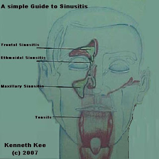
The sinuses are the air chambers in the bone behind your cheeks, eyebrows and jaw.
They produce mucus, a fluid that cleans bacteria and other particles out of the air you breathe. Tiny hairs called cilia sweep mucus out of your sinuses so it can drain out through your nose.
The paranasal sinuses are in direct communication with the nose.
The sinuses are normally sterile.

If the sinus openings may become blocked, the mucus becomes congested in the sinuses, resulting in stagnation of secretion and finally bacterial growth.

What causes sinusitis?
---------------------------

Anything that causes swelling in your sinuses or keeps the cilia from moving mucus can cause sinusitis.
This can occur because of changes in temperature or air pressure,
Using decongestant nasal sprays too much,
Smoking, and
Swimming or diving.
Some people have growths called polyps that block their sinus passages.
When sinusitis is caused by a bacterial or viral infection, you get a sinus infection.

Sinus infections sometimes occur after you've had a cold.
The cold virus attacks the lining of your sinuses, causing them to swell and become narrow.
Your body responds to the virus by producing more mucus, but it gets blocked in your swollen sinuses.
This built-up mucus makes a good place for bacteria to grow.
The bacteria can cause a sinus infection.

Acute sinusitis is usually bacterial in origin.
Haemophilus influenzae and Streptococcus pneumoniae are the organisms most commonly found in adults.
In children, similar organisms are seen, with the addition of Moraxella catarrhalis.
In older children and young adults, Staphylococcus aureus is an occasional finding.
In systemically impaired hosts, Candida, Aspergillus, and Phycomycetes may be the cause.

Risk factors include the following: diabetes mellitus, cancer, hepatic disease, renal failure, burns, extreme malnutrition, and immunosuppressive diseases.

What are the signs of acute sinusitis?
-------------------------------------------


Presentation of sinusitis is often nonspecific.

Patients may present with a persistent cold.
A cold that starts to get better and then gets worse may be a sign of acute sinusitis.

Pain or pressure in some areas of the face (forehead, cheeks or between the eyes) is often a sign of blocked sinus drainage and can be a sign of acute sinusitis.

Pain in your forehead that starts when you lean forward can also be a sign.

Other symptoms may include a stuffy nose.

Some patients complain of dental pain or alteration in smell.

Fever is seen in fewer than 2% of individuals with sinusitis.

Facial tenderness to palpation is present.

Complete opacification of sinus on transillumination is present.

An X-ray of the paranasal sinuses usually confirms the presence of sinusitis as opacity in the sinuses.

How is acute sinusitis treated?
------------------------------------

Your doctor may prescribe an antibiotic.
You may take an antibiotic for 10 to 14 days, but you will usually start feeling better a couple of days after you start taking it. It is important to take this medicine exactly as your doctor tells you and to continue taking it until it is gone, even after you're feeling better.

If you have sinus pain or pressure, your doctor may prescribe or recommend a decongestant to help your sinuses drain.

Painkillers may be prescribed if there is severe pain.

How to take care of sinusitis?
-----------------------------------

1.Get plenty of rest.

2.Lying down can make your sinuses feel more congested, so try lying on the side that lets you breathe the best.

3.Drink plenty of fluids.

4.Apply moist heat by holding a warm, wet towel against your face or breathing in steam through a cloth or towel.

5.Rinse your sinus passages with a saline solution.

How is chronic sinusitis treated?
---------------------------------------

In cases where the acute sinus infection does not cleared or become chronic, a sinus washout may be necessary to remove the mucus stucked in the sinuses.

This involves syringing of antiseptic solution through a hole in the septum separating the maxillary sinuses from the nose.
Sometimes syringing of frontal sinuses can be done through a tube inserted into the sinuses.

In severe case of chronic sinusitis, endoscopy surgery may be done to strip the lining of the maxillary sinuses and clean the cavity of the sinuses.

A new therapy is the use of phage therapy where bacterial viruses are used to cause bacterila lysis in the sinuses.

Subscribe Now: Feed Icon

Clicktale

Click and bookmark these Social Networking Bookmarks

Social Bookmarking bookmark at folkd

Labels

Is the medical Guide simple enough?

Ads by Adbrite